
Motor Evoked Potentials
Discover how Melior’s unique phenotypic screening platforms can uncover the untapped value of your candidate therapeutic
Nerve conduction and/or neuromuscular function following drug treatment, injury, or as a function of genetic factors can be evaluated in a variety of ways. Stimulation of the sciatic nerve at the hip produces compound motor action potential (CMAP) responses in the plantar foot muscle that are delayed and reduced in amplitude in SOD (superoxide dismutase) knockout (SOD -/-) or transgenic (G93A) mice. Mice can be evaluated as early as 5 weeks of age and can be tested weekly or biweekly.
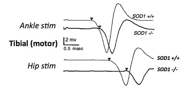
Responses in the plantar foot muscle to stimulation of the sciatic nerve at the hip and the tibial nerve at the ankle in SOD knockout (-/-) versus wild type (+/+) mice. Note the relatively delayed response latency (indicating slower conduction) and reduced muscle response amplitude in the knockout mice. (Flood et al, 1999).
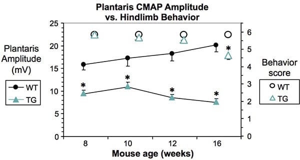
Plantar foot muscle responses evoked by stimulation of the tibial nerve were recorded from G93A SOD transgenic and non-transgenic control mice biweekly from 8 to 16 weeks. A significant difference was seen at all time points. Hindlimb behavior scores, however, were not different until 16 weeks.
* P< 0.05, unpaired t-test; N= 10-12/group.
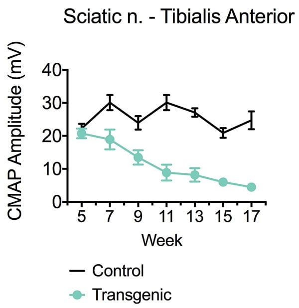
Responses in the tibialis anterior to sciatic nerve stimulation in G93A SOD transgenic versus non-transgenic control mice weekly from 5 to 17 weeks. A significant difference was seen as early as 7 weeks.
* P< 0.05, unpaired t-test; N= 12/group.
 Interested in evaluating Motor Evoked Potential?
If you are interested in learning more about Motor Evoked Potentials, please contact us to start the conversation today!