
Pulmonary Edema Model
Discover how Melior’s unique phenotypic screening platforms can uncover the untapped value of your candidate therapeutic
Pulmonary edema is caused by leakiness or extravasation of pulmonary blood vessels that can be associated with a number of pulmonary disease conditions including COVID-19. Fluid laden pulmonary alveoli have poor oxygen exchange rates and are associated with reduced respiratory function. Patients experience lower oxygen saturation levels (SpO2) leading to Acute Respiratory Distress Syndrome (ARDS) causing multiple organ failure and death. In this regard, pulmonary edema is a critical component among a sequence of events in respiratory diseases such as COVID-19 that lead to death. Therapeutic agents that can prevent or mitigate pulmonary edema may be life-saving.
Melior uses a pulmonary edema model that recapitulates important aspects of the human condition. Specifically, systemic inflammation is induced with a bolus administration of lipopolysaccharide (LPS) systemically (IP) or intranasally (IN) . The ensuing pulmonary edema can be monitored by measuring lung wet weight / dry weight ratio or by systemic administration of Evan’s Blue dye followed by dye extraction from lung tissue.
The study outlined below illustrates the pulmonary edema model in C57BL/6 mice created by an LPS-induced cytokine storm. Dexamethasone, which attenuates this cytokine storm, significantly reduces the pulmonary edema.
Ready to get started or looking for a custom model?
Contact us today for more information about our bespoke research models and to discuss how we can help you answer your unique research questions.
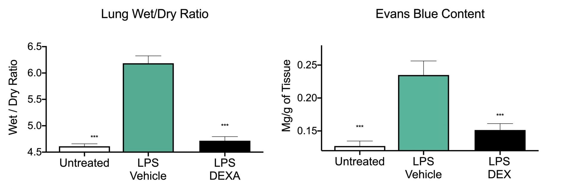
Mouse pulmonary edema model. C57BL/6 mice (10 weeks of age) were administered a bolus intraperitoneal injection of LPS (20 mg/kg). Evans Blue Dye (EBD) was administered by intravenous injection 5.5 hours after LPS administration. Six hours after LPS administration animals were sacrificed. EBD was extracted with formamide and density measured at 610 nm, the amount was quantified against a standard curve. The weight of the other freshly dissected lung was measured and compared to lung weight after a 48 hour desiccation procedure. Data are mean± SEM; ***p< 0.001 compared to LPS – Vehicle group (N= 8).
The mouse pulmonary edema model is typically run as an acute model (one/two day day study, 6-48 hrs after LPS administration) with a single administration of test article. In addition to monitoring measures of edema it is also common to incorporate measures of cytokines such as IL-6, TNF-α, and MCP-1.
Frequently Asked Questions
Evan’s Blue is administered via IV 30 mins prior to euthanasia. Evan’s blue is a large dye molecule and has high affinity for serum albumin. Evan’s blue is used to assess the permeability of the lungs. In a normal lung, no Evan’s blue should be seen. However, in pulmonary edema, the leakiness of the pulmonary system results in Evan’s blue being found in the lungs.
This model is typically run as an acute model where animals are euthanized 6-48 hours after LPS administration with a single administration of test article.
Absolutely! We often measure IL-6, TNFa, and MCP-1 in this model. We can measure other cytokines, if requested. Make sure to talk to our scientists to make sure your cytokine of interest plays a role in this model.
Synonyms: ARDS, COVID-19, Respiratory Distress
 Interested in running a Pulmonary Edema study?
If you are interested in learning more about the rodent pulmonary edema model please contact us to start the conversation today!