
C-fiber Pain Reflex
Discover how Melior’s unique phenotypic screening platforms can uncover the untapped value of your candidate therapeutic
C-fibers are small unmyelinated fibers transmitting diffuse pain signals from the periphery to the spinal cord. Larger and faster myelinated A∂ & Aβ fibers transmit pain and touch information.
C-fiber pain activity can be selectively evaluated by stimulating the footpad and recording reflex activity in the peroneal muscle between 150 and 400 msec following the stimulus. Activity in this pathway can be recorded for several hours allowing within-animal determination of baseline, test article, and drug interaction effects.
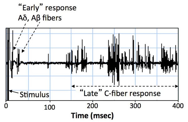
Stimulation of the ventral foot pad produces a reflex activation of the peroneus longus muscle consisting of early Aδ and Aβ nerve fiber responses, followed by the unmyelinated C-fiber response beginning around 150 msec.
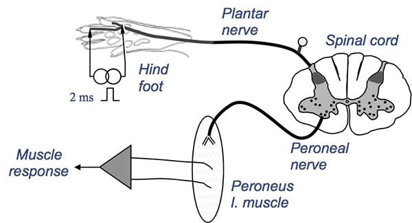
The C-fiber response (CFR) is recorded from the peroneus longus muscle during continuous stimulation of the ventral foot pad at 6 sec intervals
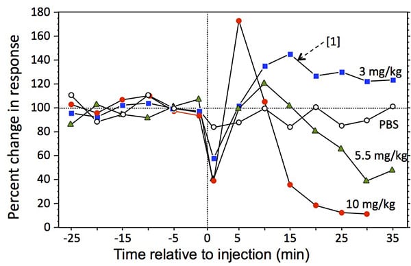
Dose-related effect of morphine on CFR. Morphine was administered subcutaneously at time 0; N=3 rats per curve. The CFR was completely inhibited at 10 mg/kg for approximately 30 min. [1] Note the increased response at 3 mg/kg – this is normal and presumed to result from greater supra-spinal dis-inhibition relative to spinal inhibition at this dose. The effect of morphine on the CFR is reversed by the opioid antagonist naloxone (not shown).
 Interested in running a C-fiber Pain Reflex study?
If you are interested in learning more about the C-fiber Pain Reflex, please contact us to start the conversation today!