
Cortical Sensory Evoked Potentials
Discover how Melior’s unique phenotypic screening platforms can uncover the untapped value of your candidate therapeutic
Somatosensory (tibial n., auditory, etc.) evoked or event-related potentials can be recorded at various locations in the brains of rats and mice using surface or depth electrodes. These potentials can be used to monitor the effects of drugs on attentional and cognitive functions.
Auditory evoked potentials have been recorded from the hippocampus, for example, to evaluate the effects of drugs on attention. A topographic map of EEG responses to auditory clicks shows functional localization of responses across the cortex.
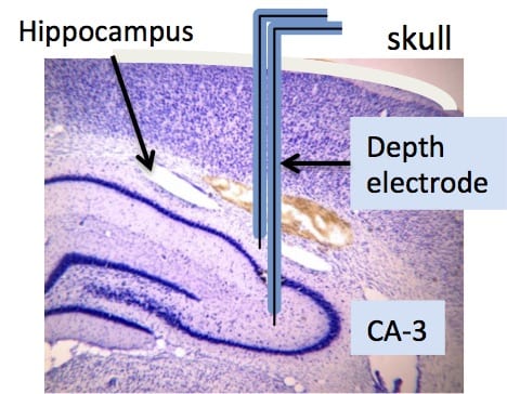
Illustration of bipolar depth electrode implanted into the CA3 region of the hippocampus.

Hippocampal evoked responses to an auditory conditioning tone (blue) followed 0.5 sec later by a test tone (red) (tones = 3 kHz x 10 msec duration). Tone-pairs were delivered 10 sec apart; N= 24. The test tone amplitude (P1 – N1) was reduced by approximately 50% compared to the conditioning tone.
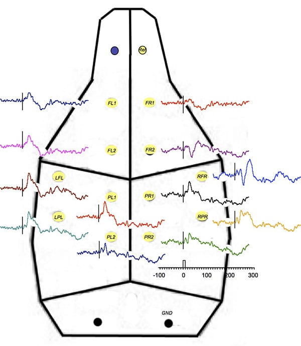
This figure shows average evoked responses from a rat implanted with 12 separate chronic supra-dural electrodes using a common reference over the olfactory bulb. Responses to auditory clicks (75 dB, 10 msec, 3 kHz, 11 sec ISI, N= 32) from a speaker above the animal are shown adjacent to electrode position. The largest response at location “RFR” (right fronto-parietal region) was above the auditory cortex. Note the different shapes of potentials around this location indicating localized surface responses. Time scale on the bottom axis in msec; tone at time 0.
 Interested in evaluating Cortical Sensory Evoked Potentials in mice or rats?
If you are interested in learning more about the Cortical Sensory Evoked Potentials, please contact us to start the conversation today!