Human Ovarian Cancer Model / SK-OV-3
Discover how Melior’s unique phenotypic screening platforms can uncover the untapped value of your candidate therapeutic
Ovarian cancer is the eighth most common cancer among women worldwide, with an estimated 313,959 new cases and 207,252 deaths in 2020. It is more common in developed countries, where it is the seventh most common cancer in women. It has a relatively low survival rate, especially in advanced stages making it the fifth most common cause of cancer-related death among women
Surgery is the main treatment for ovarian cancer where, for early-stage ovarian cancer, it can be curative. In advanced-stage ovarian cancer, surgery is usually followed by chemotherapy or other more advanced therapeutic strategies. For example. several PARP inhibitors have been approved for the treatment of ovarian cancer, including olaparib, niraparib, and rucaparib. While immune checkpoint inhibitors (ICIs) have provided revolutionary advances for some forms of cancer, unfortunately clinical trials to date evaluating ICIs (PD1/PD-L1) in patients with relapsed ovarian cancer have been disappointing. Combining ICIs with chemotherapy, anti-VEGF therapy, or PARP inhibitors improved response rates and survival in spite of a worse safety profile. Therefore, there remains high unmet medical need for improved therapeutic agents. Melior’s animal model of ovarian cancer is an important tool towards this goal.
SK-OV-3 cells are a well-established cell line that has been widely used as a model of ovarian cancer. They were originally derived from the ascites fluid of a 64-year-old Caucasian female with ovarian adenocarcinoma. SK-OV-3 cells grow rapidly in culture and can reach confluence within 3-4 days with a doubling time of approximately 39 hours. They are known to carry mutations in the TP53 and PIK3CA genes, which are commonly found in human ovarian cancers. Also, they have a near-diploid karyotype. SK-OV-3 cells express several markers associated with ovarian cancer, including CA-125 and HE4 and are sensitive to chemotherapy drugs commonly used in the treatment of ovarian cancer, such as paclitaxel and cisplatin.
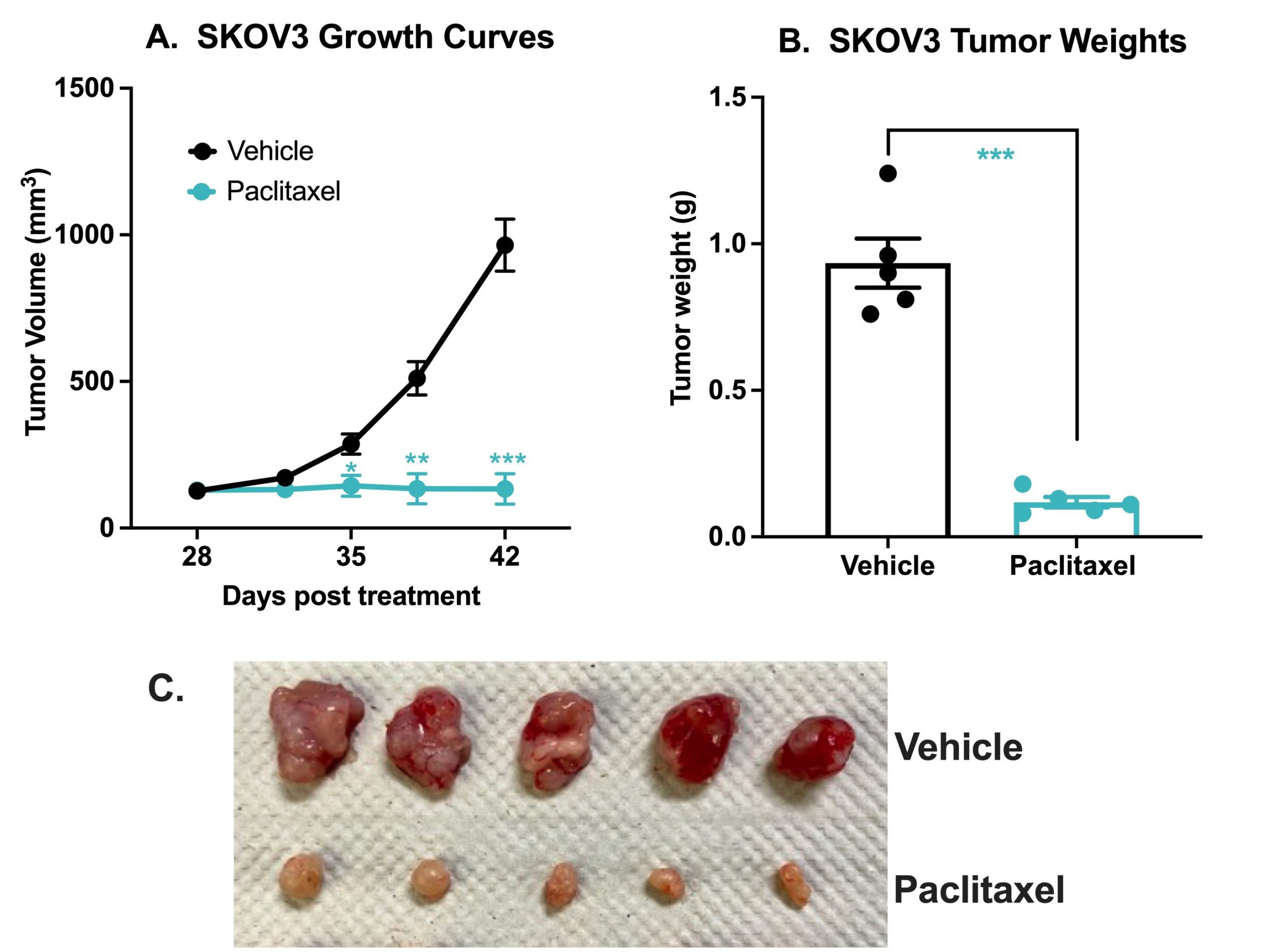
Human ovaria cancer SK-OV-3 xenograft model. 3×10⁶ SK-OV-3 cells were subcutaneously injected into the rear flank of nude mice. Once the tumor size reached ~100 mm3 (Day 28) mice were randomized into vehicle control group (treated with normal saline) and paclitaxel (20 mg/kg IP once/week). The growth of tumor was monitored twice per week using calipers (A). At the end of the study (Day 42) animals were sacrificed, tumors excised, and weighed (B, C). Data area mean +/- SEM. N=5. * P< 0.05, ** P < 0.01, *** P< 0.001 by Student’s t-test.
Melior can initiate your SK-OV-3 ovarian cancer model study with very short lead times and with bespoke study design to suit your needs. Including time to establish tumor-bearing mice (3-4 weeks) and typical treatment times (2-3 weeks) these studies normally run for approximately 6-7 weeks..
References
- National Cancer Institute. Ovarian Cancer Treatment (PDQ®)–Patient Version. Available at: https://www.cancer.gov/types/ovarian/patient/ovarian-epithelial-treatment-pdq


 Interested in running a human ovarian cancer model study?