
The PANC-1 Xenograft Model for Pancreatic Cancer
Test your pancreatic cancer therapeutics reliably and rapidly in a translatable pancreatic cancer model.
Pancreatic cancer is an aggressive malignancy arising from the pancreas, often diagnosed late due to vague symptoms. It has poor prognosis, limited treatment options, frequent metastasis, and remains one of the deadliest cancers despite advances in surgery, chemotherapy, and immunotherapy.
The PANC-1 xenograft mouse model is a valuable platform for evaluating potential therapeutics targeting pancreatic cancer, aiding in the advancement of treatment strategies and improving patient outcomes.
A translatable pancreatic tumor model platform
The PANC-1 xenograft model is a widely used preclinical system for studying pancreatic cancer biology and therapeutic responses. PANC-1 is a human pancreatic ductal adenocarcinoma cell line, originally derived from a pancreatic carcinoma in a 56-year-old patient. When implanted into immunodeficient mice, typically either subcutaneously or orthotopically into the pancreas, these cells form tumors that recapitulate several features of human pancreatic cancer, including aggressive growth, stromal interactions, and relative resistance to therapy. The subcutaneous model offers easy monitoring of tumor volume, while the orthotopic model more closely mimics the tumor microenvironment and metastatic potential. PANC-1 xenografts are particularly valued for evaluating novel chemotherapies, targeted therapies, and combination regimens, as well as for studying tumor biology, epithelial–mesenchymal transition, and mechanisms of drug resistance. This model provides a reproducible and clinically relevant platform to advance translational research in pancreatic cancer.
Advantages of the PANC-1 Xenograft Model
- Can be grown orthotopically to faithfully re-capitulate 3D environment
- Most commonly used human pancreatic cell line.
- Reflect clinical treatment challengeswith demonstrated chemotherapy resistance.
Customize your lung cancer research with our bespoke services
Our PANC-1 xenograft model can be prepared in 2 to 3 weeks, with results available after 2-3 months of treatment.
Contact us for more information on our additional and individualized services, including IVIS imaging, PK studies, and metabolic analyses.


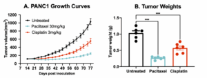
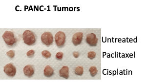
Human pancreatic cancer PANC-1 xenograft model. 10×106 PANC-1 cells were subcutaneously injected into the rear flank of NSG mice. Once the tumor size reached ~100mm3 (Day 14), mice were randomized into 3 groups (n=6 each): untreated control, paclitaxel (30 mg/kg, IP weekly) and cisplatin (3 mg/kg, IP weekly). Tumor volume was monitored 2x weekly using calipers (A). At the end of the study (Day 44), animals were sacrificed, tumors excised and weighted (B,C). Data are mean ± SEM; *** p<0.001.
Did you know the A549 xenograft model is complementary to our syngeneic LLC model?
Melior makes syngeneic lung cancer models that use mouse rather than human lung cancer cell lines opening up the possibility of immune therapy testing. Our Lewis Lung Cancer (LLC) syngeneic model complements the human A549 xenograft model, and both are models of NSCLC.
Publications
Frequently Asked Questions
PANC-1 cells were originally derived from a pancreatic carcinoma in a 56-year-old patient
The PANC-1 xenograft model is the most commonly used pancreatic xenograft model thereby allowing study results to more easily be compared to literature results.
Melior can either implant PANC-1 cell subcutaneously, allowing for relatively straight forward measurement of tumor volume, or implant cell orthotopically into the pancreas. This has the advantage of provide a more natural tumor environment and tumor volume is measured using bioluminescent imaging.
Citations
Lieber, M., Mazzetta, J., Nelson-Rees, W. A., Kaplan, M., & Todaro, G. J. (1975). Establishment of a continuous tumor-cell line (PANC-1) from a human carcinoma of the exocrine pancreas. International Journal of Cancer, 15(5), 741–747. https://doi.org/10.1002/ijc.2910150505.
Deer, E. L., González-Hernández, J., Coursen, J. D., Shea, J. E., Ngatia, J., Scaife, C. L., Firpo, M. A., & Mulvihill, S. J. (2010). Phenotype and genotype of pancreatic cancer cell lines. Pancreas, 39(4), 425–435. https://doi.org/10.1097/MPA.0b013e3181c15963.
Kim, M. P., Evans, D. B., Wang, H., Abbruzzese, J. L., Fleming, J. B., & Gallick, G. E. (2009). Generation of orthotopic and heterotopic human pancreatic cancer xenografts in immunodeficient mice. Nature Protocols, 4(11), 1670–1680. https://doi.org/10.1038/nprot.2009.171.
Delitto, D., Judge, S. M., Delitto, A. E., Nosacka, R. L., Rocha, F. G., DiVita, B. B., Gerber, M. H., George, T. J., Behrns, K. E., Hughes, S. J., Wallet, S. M., Judge, A. R., & Trevino, J. G. (2016). Human pancreatic cancer xenografts recapitulate key aspects of cancer cachexia. Oncotarget, 8(1), 1177–1189. https://doi.org/10.18632/oncotarget.13593.
Garcia, P. L., Miller, A. L., & Yoon, K. J. (2020). Patient-derived xenograft models of pancreatic cancer: Overview and comparison with other types of models. Cancers, 12(5), 1327. https://doi.org/10.3390/cancers12051327.
American Type Culture Collection. (n.d.). PANC-1 (CRL-1469) [Cell line]. ATCC. Retrieved September 24, 2025, from https://www.atcc.org/products/crl-1469. https://www.atcc.org
Cellosaurus. (n.d.). PANC-1 (CVCL_0480). Retrieved September 24, 2025, from https://www.cellosaurus.org/CVCL_0480.